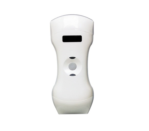
Sonography is a non-invasive, painless procedure where you can use a portable ultrasound machine. It uses high-frequency sound waves called ultrasound waves to produce images of organs, soft tissues, blood vessels, and blood flow, from inside the body. These images are used for medical analysis as it helps doctors gain insights into the inner workings of the body. It’s also known for being:
- Safe
- Radiation free
- Affordable
Variations of ultrasound include;
- Doppler Ultrasound- to measure and visualize blood flow in the heart and blood vessels.
- Elastography- to differentiate tumors from healthy tissue.
- Bone sonography- to determine bone density
- Therapeutic ultrasound- to heat or break up tissue
- High-Intensity Focused Ultrasound (HIFU)- to destroy or modify abnormal tissue in the body without opening the skin
IMPORTANCE
- An ultrasound can provide very important diagnostic information about a developing baby, including confirming the pregnancy and gestational age; checking for multiple pregnancies, congenital anomalies, and or problems with the placenta; monitoring fetal growth, and the level of amniotic fluid.
- Monitor fetal position
- Diagnostics
- Used during medical procedures. For example, ultrasound imaging can help doctors during procedures such as needle biopsies. These require the doctor to remove tissue from a very precise area inside the body for testing in a lab.
- Therapeutic applications. You can sometimes use ultrasounds to detect and treat soft-tissue injuries.
Types of Ultrasounds
The different kinds of ultrasounds include transvaginal ultrasound, 3-D ultrasound, 4-D ultrasound, or fetal echocardiography. There are additional types of ultrasounds that look at specific organs in the abdomen like the liver or vascular system. Most ultrasounds take approximately 30-45 minutes to complete, and after an ultrasound, patients receive the results within days.
Ultrasound is sometimes used in the diagnosis of problems after the delivery of the baby. It is not as useful as MRI or CT imaging to detect the presence or type of hypoxic-ischemic injury in the newborn brain. It is very useful in the diagnosis of brain hemorrhage and to do an initial assessment of brain anatomy to determine if there are any changes to the anatomy suggestive of prior insult.
1 in 12 children in Africa dies before turning 5. More than 430 women die each day from preventable causes related to pregnancy and childbirth according to WHO. To help save lives, some companies have started tapping new technologies that can diagnose health conditions and diseases more efficiently and accurately than current practices using standard equipment. One such technology is the M-scan which comes as a package containing an M-scan ultrasound package that includes a probe tablet and a bag with an ultrasound charger which costs $2,500. Four medical entrepreneurs in Uganda invented this type of technology.
How the Portable Ultrasound Machine (M-scan) Works.
The M-scan can work with a laptop, tablet, or smartphone to detect the factors of maternal mortality among pregnant mothers in socially limited settings. This type of technology would help in making sure that mothers don’t die due to factors that could be detected easily by an ultrasound.
The new invention is a valuable asset in prenatal and antenatal care for mothers who can’t access larger health facilities.
WHO recommends that women who have at least four antenatal visits to detect any complications in pregnancies. Many in rural areas do not have the means or access to facilities to undertake even a single visit. One ultrasound scan before 24 weeks’ gestation is crucial to estimate gestational age, improve detection of fetal anomalies, and multiple pregnancies. It can also reduce the induction of labor for post-term pregnancy, and improve a woman’s pregnancy experience.

